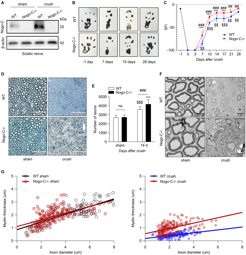
FIGURE 2.
Nogo-C knockout promotes sciatic nerve regeneration. (A) Western blot shows the Nogo-C protein level in sciatic nerves from WT and Nogo-C–/– mice. Nogo-C–/– mice show no significant expression of Nogo-C in the sham and crush groups. (B,C) Sciatic Function Index between WT and Nogo-C–/– mice before (–1 day) and after sciatic nerves injury; n = 4 mice per group. (D) A typical example of semi-thin sections of WT and Nogo-C–/– tibial nerves 2 weeks after crush injury and sham; scale bar = 20 μm. (E) Mean number of axons in tibial nerves; n = 6 mice per group. (F) Representative Toluidine blue-stained ultrathin section of WT and Nogo-C–/– tibial nerves 2 weeks after crush injury and sham; scale bar = 5 μm. (G) Myelin thickness relative to axon diameter was quantified in cross-sections of uninjured (sham) and injured (crush) tibial nerves of four WT and four Nogo-C–/– mice 2 weeks after crush injury and illustrated as a scatter plot. 50 axons and myelin sheath were measured from each nerve and shown in the plot. There were significant differences (p < 0.001) between WT and Nogo-C–/– nerves at 2 weeks of regeneration and no difference between sham groups. $$P < 0.01, $$$P < 0.001 vs. WT sham, *P < 0.05, ***P < 0.001 vs. Nogo-C–/– sham, ##P < 0.01, ###P < 0.001 vs. WT crush.

