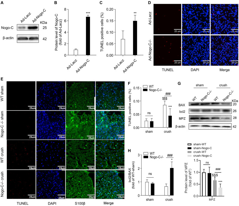
FIGURE 3.
Nogo-C regulates cardiomyocyte apoptosis. (A,B) Western blot showing Nogo-C protein expression in rat neonatal Schwann cells transfected with Ad-Nogo-C at 75 MOI or Ad-lacz for 48 h; n = 3 independent experiments. (C,D) TUNEL staining showing apoptotic rates of Schwann cells transfected with Ad-Nogo-C or Ad-lacz; n = 6 independent experiments. Scale bar = 25 μm. **P < 0.001, ***P < 0.001 vs. Ad-lacz cells. (E,F) TUNEL staining showing apoptotic cells in sham and crushed (7 days after the operation) sciatic nerves from WT and Nogo-C–/– mice. n = 4 mice. Scale bar = 25 μm. (G,H) western blot and average data showing BAX Bcl-2 and MPZ protein levels in sham and crushed sciatic nerves from WT and Nogo-C–/– mice; n = 3 mice for each group. $$$P < 0.001 vs. WT sham, *P < 0.05, ***P < 0.001 vs. Nogo-C–/– sham, ###P < 0.001 vs. WT crush.

