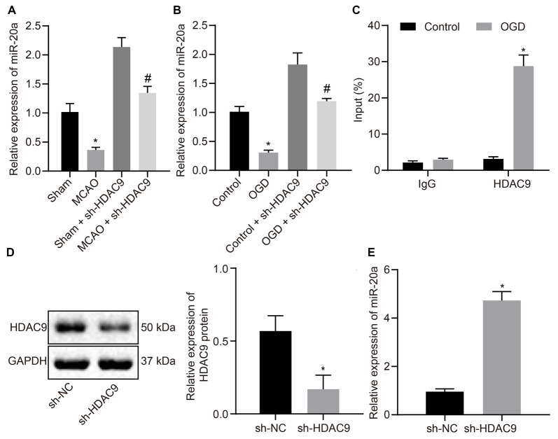
Figure 3.
HDAC9 negatively regulates miR-20a expression in vitro. (A) miR-20a expression in brain tissues of MCAO mice or MCAO mice treated with sh-HDAC9 detected by RT-qPCR (n = 8), normalized to U6. (B) miR-20a expression in OGD-treated neurons or OGD-treated neurons treated with sh-HDAC9 detected by RT-qPCR, normalized to U6. (C) HDAC9 enrichment in the miR-20a promoter region detected by ChIP. (D) Western blot analysis of HDAC9 protein in OGD-treated neurons transfected with sh-HDAC9, normalized to GAPDH. (E) miR-20a expression in OGD-treated neurons transfected with sh-HDAC9 detected by RT-qPCR, normalized to U6. *p < 0.05 vs. the sham, control, or sh-NC group. Measurement data (mean ± standard deviation) between the two groups were compared using an unpaired t-test. n = 3. #p < 0.05 vs. the sham + sh-HDAC9 group or the control + sh-HDAC9 group.

