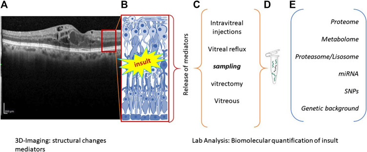
FIGURE 1.
Flowchart summarizing the main steps of precision medicine. (A), (B) Biostrumental parameters’ acquisition by computerized tomography. Representative image showing retinal layers (A) with a red square schematized in (B). Retinal insult is followed by the release of a plethora of inflammatory mediators that can be quantified according to different sampling route (C). Samples can be appropriately processed to release RNA, DNA, or proteins (D), according to different new and old generation techniques (E). The main biomarkers used as outcome indicator are listed in (E).

