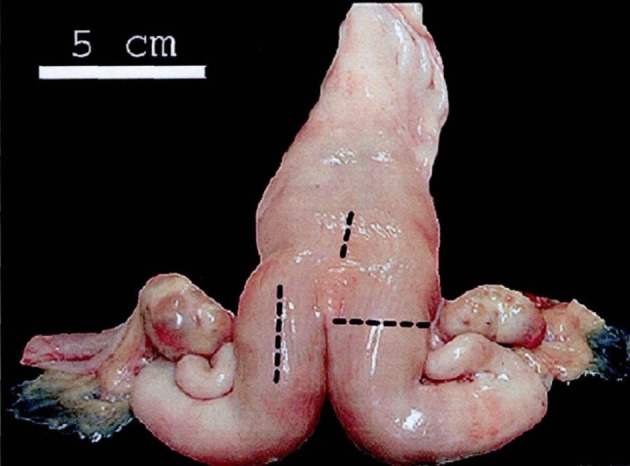
Figure 1.

Anatomical model of the bovine uterus. Black lines mark the locations where the B-mode images of the uterus were taken. One long-sectional image of each uterine horn and one long-sectional image of the cranial end of the cervix were taken using a B-mode veterinary ultrasound scanner in our study. The figure is cited from Schmauder et al. (30).
