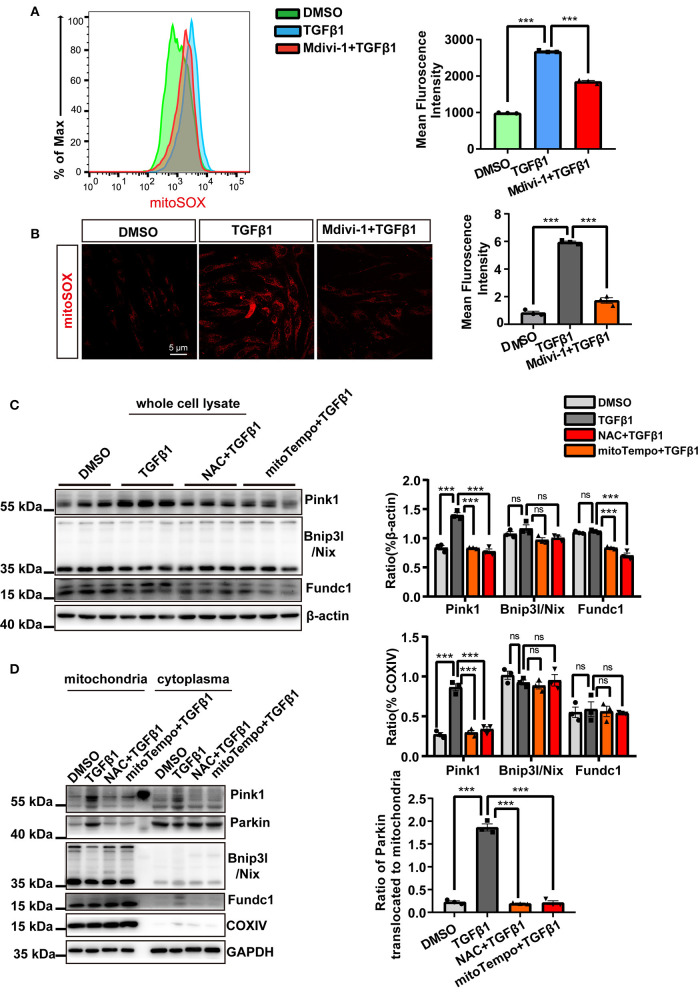
Figure 4.
Mitochondrial reactive oxygen species (mtROS) were the main instigators of mitophagy in cardiac fibroblasts (CF). mtROS was detected by MitoSOX staining and visualized by flow cytometry (A) and confocal microscopy. (B) Mean immunofluorescence intensities were calculated. (C) The expression of mitophagy-related proteins after treatment with TGF-β1 and ROS scavengers was measured in whole-cell lysates. (D) Western blotting was used to compare mitophagy-related parameters and mitochondrial parkin translocation in particular, among the different groups. COXIV was used as the loading control for the mitochondrial fraction. Data are shown as mean ± standard error of the mean (n = 3 independent cell isolations per group). Means were compared by one-way ANOVA, followed by the Student–Newman–Keuls (SNK) post hoc. ns, not significant; ***P < 0.001.

