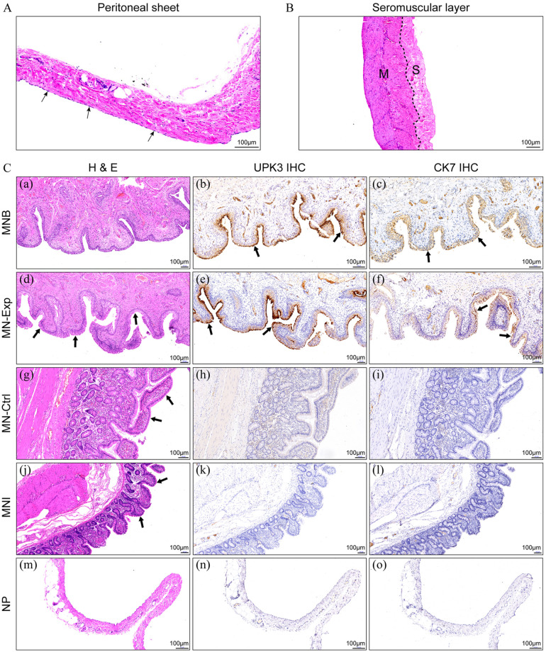
Figure 5.
A. HE staining of the peritoneal sheet. Black arrow: mesothelial cells. B. HE staining of the intact ileal seromuscular layer. M: muscle layer; S: serosa layer. C. Histopathological assessment of the neobladder constructed by both methods. Similar to what was observed in the MNB (a), HE staining revealed a thick layer of urothelium-like cells lying on the luminal surface of MN-Exp (d) (black arrow). Immunohistochemical analysis showed UPK3 and CK7 were highly expressed in the MNB (b and c) and MN-Exp (e and f) (black arrow), while no such changes were observed in the MN-Ctrl (h and i) and MNI (k and l). Compared with the MNI (j), the villi marked by the black arrow in the MN-Ctrl (g) were not atrophied 3 months after surgery. UPK3 (n) and CK7 (o) were negative on the NP (m). MNB, mucosa of the normal bladder; MNExp, mucosa of the neobladder in the experimental group; MN-Ctrl, mucosa of the neobladder in controls; MNI, mucosa of the normal ileum; NP, normal peritoneum; UPK3, uroplakin III; CK7, cytokeratin 7.

