Fig. 2.

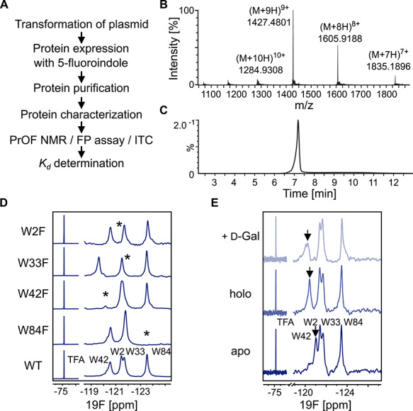
PrOF NMR of 5FW LecA. (A) General workflow for PrOF NMR with 5FW LecA. (B) Chromatogram of the LC–ESI–MS analysis of 5FW LecA. (C) ESI-MS+ spectrum of the main peak at 7.3 min [M + H]+Ca = 12826.23 Da [M + H]+found = 12831.34 Da corresponds to 5FW LecA. (D) PrOF NMR assignment of 5FW LecA WT and the mutants W84F, W42F, W33F and W2F. The tryptophanes being mutated are indicated with asterisk. All spectra were normalized and referenced to TFA. (E) PrOF NMR of 5FW LecA WT in Ca2+-free (apo, bottom) and -bound (holo, central) forms. The W42 resonance (black arrow) shifted in presence of Ca2+ and 0.5 mM d-Gal binding verifying that protein is active.
