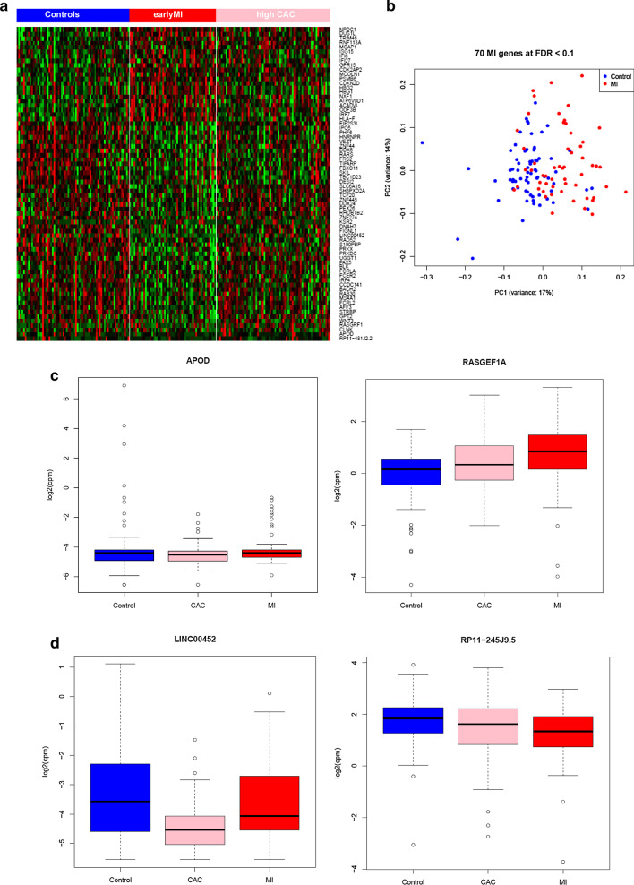
Fig. 2.
MI and CAC expression signatures. a Heatmap of 198 samples showing substantial differential expression changes across three groups (Controls, early MI, and high CAC). b Principle component analysis (PCA) of 70 MI gene signatures (68 protein-coding and 2 lincRNAs) shows a separation pattern between MI and controls. c Examples of the genes significantly associated with both MI and high CAC. The boxplot shows a low expression level in early MI and high CAC compared to controls for APOD and vice versa for RASGEF1A. d Boxplot of 2 lincRNAs that are moderately expressed in blood and associated with MI and CAC by RNA-Seq

