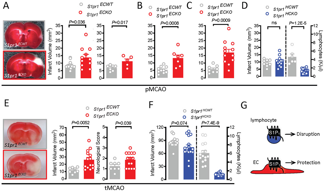
Figure 1: Endothelial S1P1 signaling limits brain injury after permanent and transient MCA occlusion.
A-C. Infarct volumes 24 (A,C) or 72 (B) hours after pMCAO in S1pr1ECKO and littermate males (A, middle panel, B) and females (A, right panel) generated by neonatal S1pr1 deletion with Pdgfb-iCreERT2 (A,B) or Cdh5-iCreERT2 (C). Left panel of A: representative images. D. Basal peripheral blood lymphocyte counts and infarct volumes 24 hours after pMCAO in male mice lacking S1P1 in hematopoietic cells (S1pr1HCKO; Vav1-Cre). E. Infarct volumes and neurological deficits 24 hours after 60 minutes tMCAO in male mice lacking S1P1 in endothelial cells (S1pr1ECKO; Cdh5-iCreERT2; adult deletion). Left panel: representative images. F. Infarct volumes 24 hours after 60 minutes tMCAO in males lacking S1P1 in hematopoietic cells (S1pr1HCKO; Mx1-Cre). Lymphocyte counts pre-MCAO in right panel. G. Schematic representation of the net cell type specific contribution of S1P1 signaling to stroke outcome. Bar graphs show mean ± SEM. Statistical significance assessed by Mann-Whitney test (A, males) or unpaired t-test (all other).

