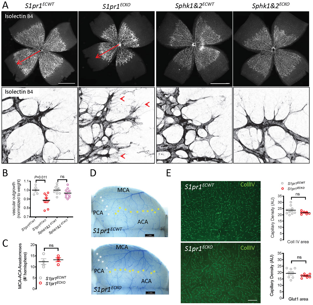
Figure 3: Loss of EC-autonomous S1P1 signaling does not impact cerebrovascular patterning.
A. Isolectin B4 staining shows vascularization of the mouse retina at postnatal day (P)5 after neonatal Pdgfb-driven S1pr1 or Sphk1&2 deletion. Note delayed expansion of the vascular network (top panel, arrow) and abundant filopodia at the vascular front (bottom panel, arrowheads) of S1pr1ECKO but not Sphk1&2ECKO retinas. Scale bar: 1 mm (top panel), 50 μm (bottom panel) B. Quantification of outgrowth of the retinal vasculature at P5, normalized to weight of pups. C,D. Collateral connections between the MCA and branches of the posterior CA (PCA, white asterisk) and of the anterior CA (ACA, yellow asterisk) extending laterally from the midline between S1pr1ECKO and littermate controls. C, quantification of ACA-MCA connections. D, representative images. E. Vascular density in the cortex of S1pr1ECKO and littermate controls assessed by collagen IV (Coll IV) or Glut1 staining. Representative images of Coll IV staining (left panels) and quantification of both (right panel), scale bar: 100 μm. Bar graphs show mean ± SEM. Statistical significance assessed by one-way ANOVA with Tukey multiple comparisons test (B) and unpaired t-test (all other).

