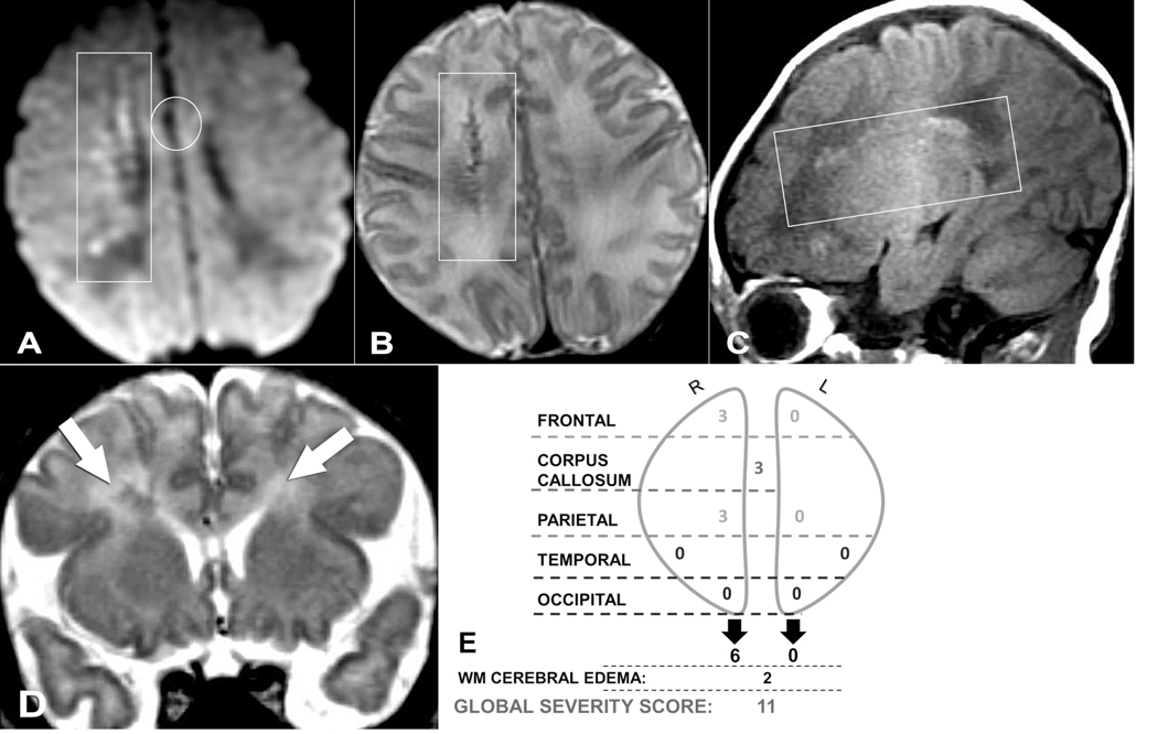
Figure 2.
(A) Axial DWI, (B) axial T2-weighted, (C) sagittal T1-weighted, and (D) coronal T2-weighted magnetic resonance images (MRIs) of a 7-day-old male neonate who presented with seizures. (E) Global severity score = 11 (second quartile). There are severe right frontal (3 points) and severe right parietal (3 points) hemorrhagic linear lesions (T1 bright and T2 dark) with restricted diffusion (+DWI) (A-C, boxes). There is restricted diffusion throughout the corpus callosum (genu shown, A, circle) (3 points) and asymmetric, bilateral white matter edema, R > L (D, arrows) (2 points). Neuropsychological testing was normal at 8 years of age, and the patient had no major neurodevelopmental or neurosensory impairment. DWI, diffusion-weighted imaging; R, right; L, left; WM, white matter.

