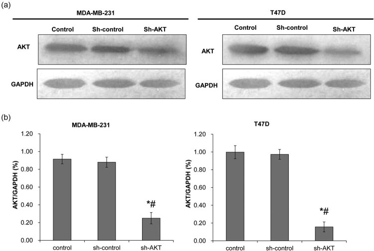
Figure 1.
Determination of lentiviral transfection efficiency. Western blot was used to detect AKT protein expression after shRNA transfection. The control group was not transfected with shRNA; the sh-control group was transfected with void vector; and the sh-AKT group was transfected with shRNA of AKT. (a) Representative Western blot results. (b) The quantified expression of AKT. Left: MDA-MB-231 cells; right: T47D cells. *P < 0.05, compared with the control group; # P < 0.05, compared with the sh-control group.

