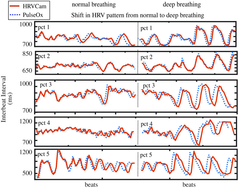
Fig. 13.
Change in HRV pattern from normal breathing to deep breathing in a participant: In the left panel, the pattern of IBI is random while the person breathes normally, and its HRV variability is lower. On the right panel, during deep breathing due to respiration sinus arrhythmia, the pattern of the IBI is sinusoidal syncing with the respiration, and HRV variability increases. We show examples of five random participants (top to down).

