Figure 3.

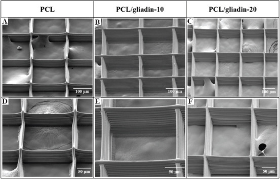
Scanning electron microscope images and their corresponding enlarged views of morphology. (A) and (D) poly(e-caprolactone) (PCL). (B) and (E) PCL/gliadin-10. (C) and (F) PCL/glaidin-20 scaffolds.

Scanning electron microscope images and their corresponding enlarged views of morphology. (A) and (D) poly(e-caprolactone) (PCL). (B) and (E) PCL/gliadin-10. (C) and (F) PCL/glaidin-20 scaffolds.