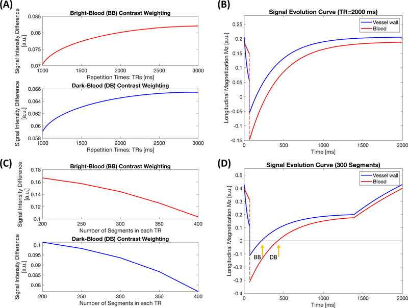
Figure 2.
Simulation results. (A) Curve of lumen-wall contrast with respect to bright-blood and dark-blood contrast weightings against various TRs with no gap until the next T2-preparation inversion recovery (T2-IR) prepared pulse. (B) Signal evolution curves of the aortic vessel wall and lumen blood with a 2000-ms repetition time. (C) Curve of lumen-wall signal difference against numbers of segments during a single readout block with TR = 2000 ms. (D) Signal evolution curves of the aortic vessel wall and lumen blood with 300 readout segments followed by a 600-ms gap until the next preparation pulse, under the circumstance of TR = 2000 ms. Two representative time points were selected for bright-blood and dark-blood contrast weightings during the simulation study.

