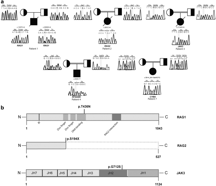
FIG. 2.
Mutation analyses of the patients. (a) Sanger sequencing results display the mutations in the patients and segregation pattern in the parents. (b) Domain architecture of the RAG1 and JAK3 proteins and locations of the missense mutations. Schematic representations of the truncated RAG2 protein compared with wild type one.

