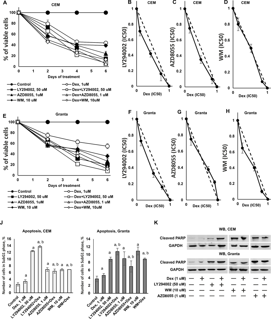
Figure 3. Synergistic and additive cytotoxic effects of PI3K/mTOR/Akt inhibitors and Dexamethasone in CEM and Granta cells.
(A-E) Cytotoxic effect was determined using MTT assay. CEM (A) and Granta (E) cells were pretreated with solvent (Control), LY294002 (50 uM), AZD8055 (1 uM) and WM (10 uM) for 6 h and treated with either solvent or glucocorticoid Dex (1 uM) for 24–144 h. (B-D) Isobologram analysis in CEM cells for the combination of LY294002 (B), AZD8055 (C), WM (D) and Dex. (F-H) Isobologram analysis in Granta cells for the combination of LY294002 (F), AZD8055 (G), WM (H) and Dex. The concentration, which resulted in 50% cell growth inhibition (IC50), is expressed as 1.0 on X and Y axis of the isobologram. Y-axis: LY294002, AZD8055 or WM (IC50); X-axis: Dex (IC50). (J, K) Apoptosis induction was evaluated by flow cytometry using PI staining (J) and by Western blot analysis of cleaved PARP level (K). GAPDH was used as loading control. The mean +/− SD was calculated for three individual samples/condition. Statistically significant differences as compared to: a-control; b-Dex (p<0.001) as and where indicated.

