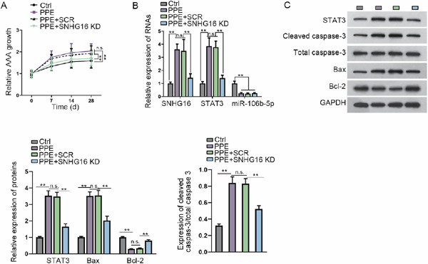
Fig. 5.

SNHG16 knockdown retards AAA formation in vivo
(A) Relative diameter of the aorta at the baseline and at 7, 14, and 28 days after aneurysm induction. AAD was assessed using B-mode ultrasound imaging. Data are expressed as growth fold change. In vivo SNHG16 knockdown was realized by site-specific antisense oligonucleotides (LNA-GapmeRs, SNHG16 KD) in order to limit aneurysm progression. (B) RT-qPCR results of the levels of SNHG16, STAT3 mRNA, and miR-106b-5p in the mice of each group. (C) Western blot results and quantification of STAT3, cleaved caspase-3, total caspase-3, Bax, and Bcl-2 in the mice of each group. **P < 0.01.
