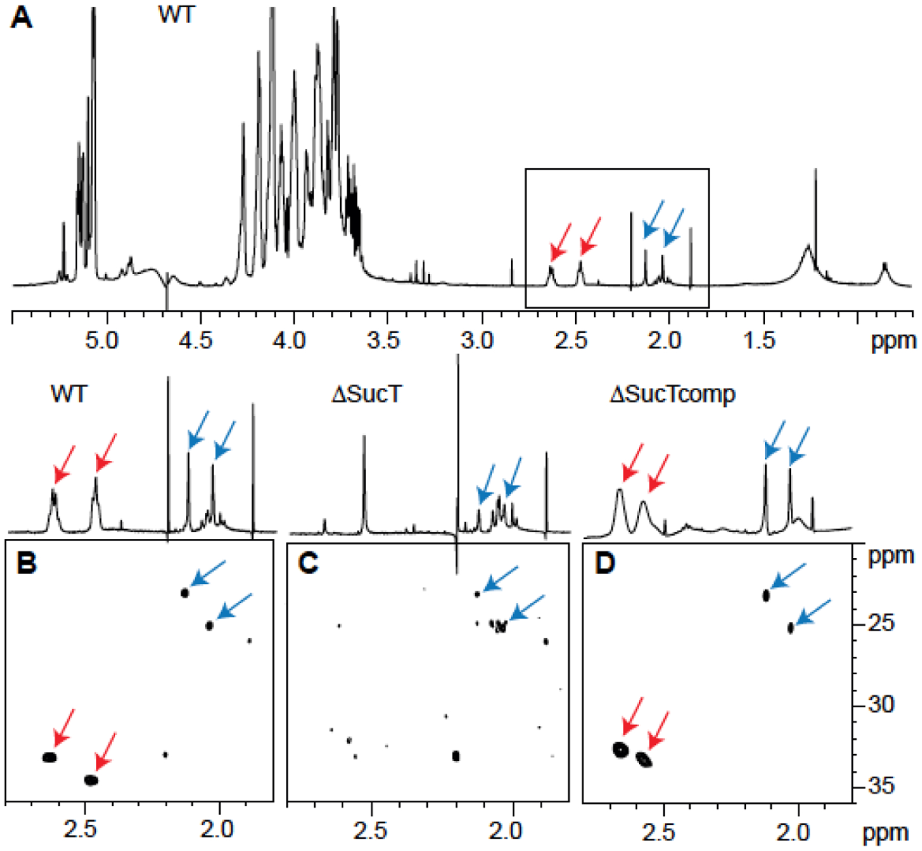
Figure 6: Presence of acetate and succinate residues on WT Mabs LAM.

NMR analysis of LAM prepared from the WT, mutant (ΔsucT), and complemented mutant (ΔsucTcomp) strains. Shown are 1D 1H (A) and expanded region (δ 1H 2.80–1.80 and δ 13C 36–20) of the 2D 1H-13C (B-D) HMQC NMR spectra. Arrows point to the signals typifying acetates (blue) and succinates (red) (see text for details).
