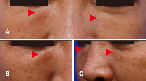
Fig. 1. (A) Skin colored nodules symmetrically distributed on both the infraorbital areas of the face. (B) Close-up view of illdemarcated, oval-shaped, and firm nodules on the right infraorbital area. The right nodule is slightly erythematous to orange colored and larger than the left one. (C) Side view of the lesions which are showing protrusion. Lesions are indicated as red arrowheads.

