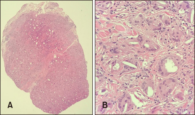
Fig. 2. (A) Histopathologic findings show multiple and scattered polymorphous foreign body reactive to fibrotic change in the dermis (H&E, ×40). (B) At higher power, non-caseating granulomas consisting of histiocytes, lymphocytes, and multinucleated giant cells with a central foreign body in the deep dermis can be visualized (H&E, ×400).

