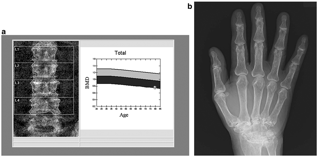
Fig. 1.

79-year-old man with a 25-year history of rheumatoid arthritis (RA). The DXA image of the lumbar spine (a) shows mild multilevel degenerative changes. A BMD of 0.762 g/cm2 was measured, which corresponds to a T-score of − 3.0. The radiograph of the right hand (b) shows advanced fusion of the carpal bones consistent with the long history of RA
