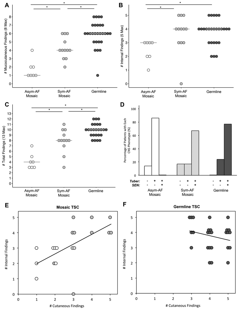
Figure 3. The Number TSC Findings and Neurological Tumor Status Differs Between Patients with Asymmetrical-Angiofibroma Mosaicism, Symmetrical-Angiofibroma Mosaicism, or Germline Disease.

(A) The number of mucocutaneous findings increased sequentially from Asymmetrical-Angiofibroma (Asym-AF) mosaicism, to Symmetrical-Angiofibroma (Sym-AF) mosaicism, to germline TSC. (B) The number of internal findings was significantly lower in patients with Asym-AF mosaicism than Sym-AF mosaicism and germline TSC. (C) The number of total findings increased sequentially from Asym-AF mosaicism, to Sym-AF mosaicism, to germline TSC. (D) The most common CNS phenotypes were tubers without SENs in Asym-AF mosaicism, and tubers with SENs in Sym-AF mosaicism and germline TSC. (E) The number of major internal and cutaneous findings correlated significantly in mosaic TSC (R=0.62, n=19, p=0.005). Those with Asym-AF mosaicism (white) clustered in the lower left quadrant whereas those with Sym-AF mosaicism (light grey) clustered in the upper right quadrant as observed in germline TSC. (F) In patients with germline TSC, the number of internal and cutaneous findings did not correlate (R=−0.24, n=26, p=0.24). These patients clustered in the upper right quadrant of the figure.
