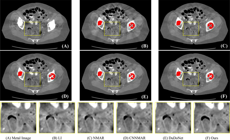
Fig. 6:
Visual comparison on CT images with real metal artifacts. The segmented metals are colored in red for better visualization. Our method effectively reduces metal artifacts and preserves the fine-grained anatomical structures. The display window of whole image is [−480 560] HU and the display window of cropped patches is [−400 300] HU.

