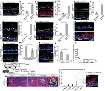
Fig. 3. Tbx3+-BCs and EPCs are maintained in proliferating plantar epidermis for homeostasis.

(A and B) Staining and quantification of PH3+-BCs in abdominal and plantar epidermis (n > 300 cells, three mice). (C) Tracing K14cre-labeled BCs. (D) Quantification of basal-to-suprabasal transition of K14cre-labeled BCs (n > 110 cells, three mice). (E and F) Staining and quantification of Tbx3+-BCs (n > 330 cells, three mice). (G and H) Staining and quantification of Adam8+-BCs (n > 100 cells, three mice). (I and J) Staining and quantification of p-ERK+–BCs (n > 120 cells, three mice). (K) Pyy staining. Arrowheads indicate Pyy+-BCs. (L and M) Staining and quantification of plantar epidermal Ki67+-BCs in control and Tbx3 cKO mice (n > 230 cells, three mice). (N and O) Staining and quantification of plantar epidermal PH3+-BCs in control and Hbegf cKO mice (n > 110 cells, three mice). (P) Tracing Tbx3cre-labeled clones. (Q) Whole-mount images of Tbx3cre-labeled clones in plantar epidermis. (R) Quantification of Tbx3cre-labeled clone survivability. (S) Distribution of the basal clone size. (T) tdTomato staining of Tbx3creERT2;R26tdTomato mice. (B, D, F, H, J, M, O, R, and S) Error bars show SEM. (B, D, F, H, J, M, and O) *P < 0.05, **P < 0.01, and ***P < 0.001, by two-tailed Student’s t test. (A, C, E, G, I, K, L, N, and T) White dashed lines indicate BMs.
