Figure 1.

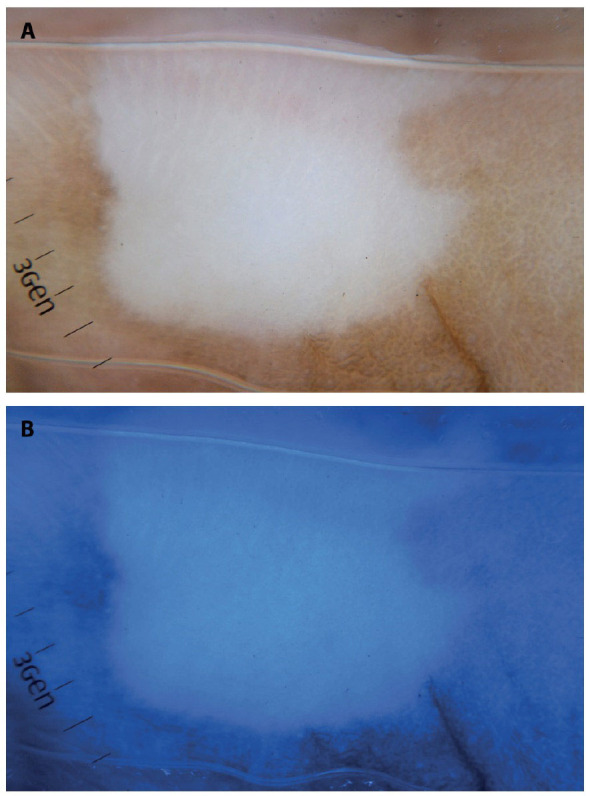
Vitiligo; stable lesion. (A) White-light and (B) blue-light dermoscopy (×10). Blue light (470 nm) delineates amelanotic vitiligo with the sharply demarcated border better than white-light dermoscopy.

Vitiligo; stable lesion. (A) White-light and (B) blue-light dermoscopy (×10). Blue light (470 nm) delineates amelanotic vitiligo with the sharply demarcated border better than white-light dermoscopy.