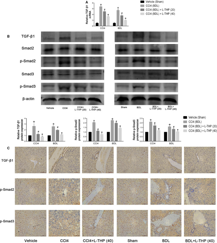
FIGURE 5.

Effects of levo‐tetrahydropalmatine on the TGF‐β1/Smad pathway in liver fibrosis: (A) The PCR analysis. The mRNA levels of TGF‐β1were significantly down‐regulated by L‐THP. Data are expressed as means ± SD (n = 3,+ P < .05 for CCl4 or BDL vs Vehicle or Sham; #P for CCl4+L‐THP (20) or BDL+L‐THP (20) versus CCl4 or BDL; *P for CCl4 +L‐THP (40) or BDL+L‐THP (40) vs CCl4 +L‐THP (20) or BDL+L‐THP (20)). B, Western blot and quantitative analysis. L‐THP treatment significantly reduced the protein expressions of TGF‐β1, p‐Smad2 and p‐Smad3. Data are expressed as means ± SD (n = 3, + P < .05 for CCl4 or BDL vs Vehicle or Sham; #P for CCl4+L‐THP (20) or BDL+L‐THP (20) vs CCl4 or BDL; *P for CCl4 +L‐THP (40) or BDL+L‐THP (40) vs CCl4 +L‐THP (20) or BDL+L‐THP (20)). C, Immunohistochemical staining indicated that the increased protein expressions of TGF‐β1, p‐Smad2 and p‐Smad3 in CCl4 and BDL groups were suppressed by 40 mg/kg L‐THP treatment (original magnification: ×400)
