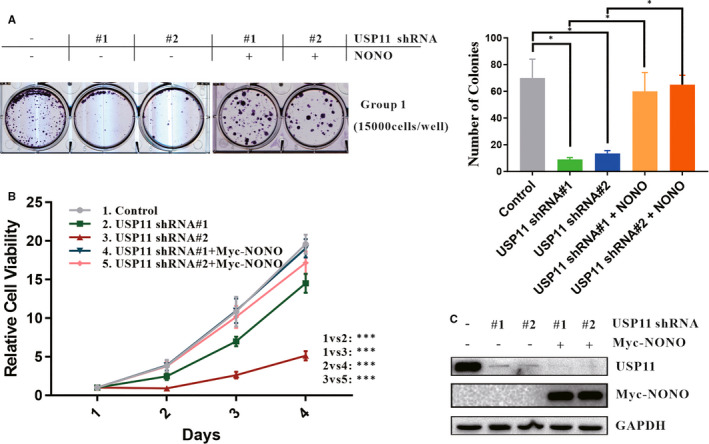
FIGURE 5.

USP11 promotes melanoma cell proliferation via NONO. A, Colony formation assay was performed. A375 cells infected with lentivirus USP11 shRNA and transfected with the indicated vectors, were seeded with density 15 000 cells per well. 24 h later, A375 cells were subcultured and selected using G418 (200 µg/mL), and surviving colonies were counted 2 weeks later. Colonies were visualized and quantified. Relative cell viability was summarized from three independent experiments and was presented on the right. Statistical significance was determined by a two‐tailed, unpaired Student's t test. Data represent the mean (±SD) of three independent experiments (*P ≤ 0.05). B, A375 was infected with USP11‐specific shRNA #1 or #2 and then introduced with Myc‐NONO plasmid. CCK‐8 assays were performed to check relative cell viability at the indicated time points. C, A375 cells used in colony formation assay and CCK‐8 assays were lysed and analysed using Western blotting
