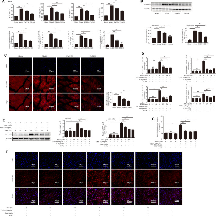
FIGURE 4.

FMN suppressed the expression of inflammation and myostatin in the muscle of CKD rats and in the TNF‐α‐induced C2C12 myotubes. (A) Levels of inflammatory markers CRP, TNF‐α, IL‐6 and IL‐1β in serum and muscle were detected by ELISA (n = 8/group) and qPCR (n = 3/group). (B) The protein and mRNA levels of myostatin were analysed in the gastrocnemius muscle (n = 3/group). (C) Myostatin expression in tibialis anterior muscles determined by (200×) immunofluorescence staining with anti‐myostatin (red) and nuclei was detected by DAPI staining (blue). The relative fluorescence intensity of myostatin was compared between the groups. (D) The levels of CRP, TNF‐α, IL‐6 and IL‐1β in C2C12 myotubes were determined by qPCR (n = 3/group). (E) The protein and mRNA levels of myostatin analysed with Western blotting and qPCR (n = 3/group). (F) Myostatin expression determined with immunofluorescence staining (200×) with anti‐myostatin (red) and nuclei detected with DAPI staining (blue). (G) The relative fluorescence intensity of myostatin was compared between the groups. *P < .05, **P < .01
