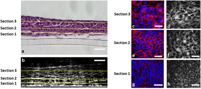
Figure 2.
Histological, immunocytochemical, and FF-OCT images of multilayered cultivated cell sheets. (a) After the complete culture and formation of mCLESs, haematoxylin and eosin (H&E) staining of the cross-sectional cell product revealed multilayered cell products. (b) FF-OCT cross-sectional image of mCLESs. (a,b) Sections 1, 2, and 3 represent the basal cell layer at 5 µm, wing cell layer at 20 µm, and superficial squamous epithelial layer at 40 µm above the bottom of the culture plate, respectively. (c,e,g) En face images of whole-mount immunocytochemical staining of mCLESs obtained from in vitro confocal microscopy at Section 1 (g), Section 2 (e), and Section 3 (c). Red: Actin. Blue: Hoechst 33,258 for the nucleus. (d,f,h) FF-OCT en face images of mCLESs at Section 1 (h), Section 2 (f) and Section 3 (d). Scale bar = 20 µm. mCLESs = multilayer cultivated limbal epithelial sheets.

