Figure 5.
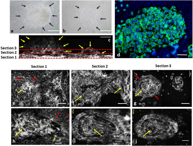
The immunocytochemical, 2D cross-sectional, and en face FF-OCT images of a complex culture system with different types of cells mixed. (a,b) Phase contrast inverted microscopy demonstrated that two DACNs grew on a sheet of epithelial cells. (c) FF-OCT cross-sectional images of the two DACNs. The height of 1 DACN was 150 μm. (d) Fluorescein microscopy en face images of the immunocytochemical staining of DACNs (Green: beta-III tubulin; Blue: Hoechst 33258 for nucleus). The staining pattern demonstrated two cell types inside and outside DACNs. (e–g) FF-OCT en face image sequence of the culture cells. Section 1 (e,h), section 2 (f,i), and section 3 (g,j) represent the cell at 5 µm, 20 µm, and 50 µm above the bottom of the culture plate. (e,f,g) Sequential images in an area with 2 DACNs illustrated in a single field. Red arrows: epithelial type of cells spread at the bottom of the culture plate, and covered the surface of DACNs. Yellow arrows: cells in the centre of DACNs exhibited different morphologies compared with epithelial cells. (h,I,j) Sequential images from an area with another DACN in a single field. Red arrows: epithelial cells spread at the bottom of the culture plate and covered the surface of DACNs. Yellow arrows: cells in the centre of DACNs exhibited different morphologies to epithelial cells. (a,b) scale bar = 200 μm, (c-j), scale bar = 50 μm. DACN: directly adherent colonies of neuron-like cells.

