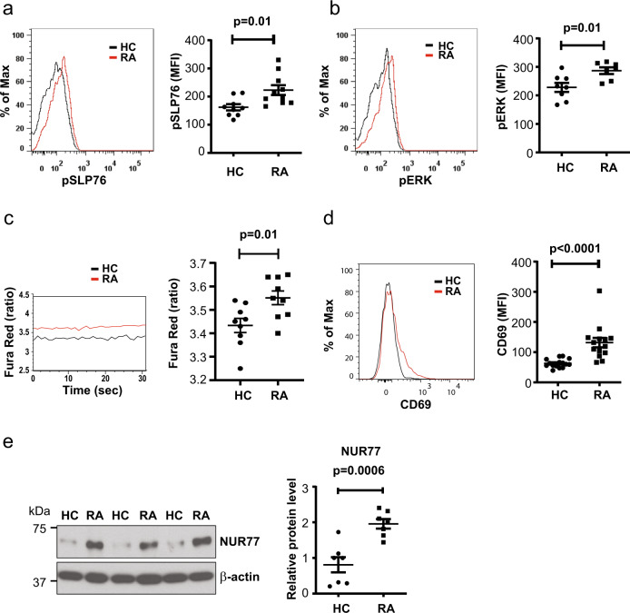
Fig. 1. Constitutively activated TCR signaling in peripheral naive RA CD4+ T cells.
Peripheral blood mononuclear cells (PBMCs) were analyzed by flow cytometry directly ex vivo without prior in vitro stimulation. Samples of rheumatoid arthritis (RA) patients and healthy controls (HCs) were always run in parallel. a Representative histograms and mean fluorescence intensity (MFI) of phosphorylated SLP76 (Y128) in gated CD3+CD4+CD45RA+CD62L+ naive CD4+ T cells from HC (n = 9) and RA (n = 10) patients are shown. Gating strategies for naive CD4+ T cells is shown in Supplementary Fig. 1a. b Representative histograms and MFI of phosphorylated ERK (Thr202/Tyr204) in CD3+CD4+CD45RA+CD62L+ naive CD4+ T cells from HC (n = 8) and RA (n = 7) patients. c Representative tracing of Fura Red ratios (left) and shifts in Fura Red fluorescence (at 406 and 532 nm) in naive CD4+ T cells from HC (n = 9) and RA (n = 9) patients (right). d Representative histograms of CD69 expression in CD3+CD4+CD45RA+CD62L+ naive T cells (left) and data from HC (n = 15) and RA (n = 14) patients (right). e NUR77 expression in isolated naive CD4+ T cells shown as representative immunoblots (left) and plots of relative densities from HC (n = 7) and RA (n = 7) normalized to β-actin (right). Uncropped Western blots in Supplementary Fig. 9. All data are presented as dot plots with mean ± SEM. All statistical analyses were performed with unpaired two-tailed Student’s t test. Source data are provided as a Source Data file.

