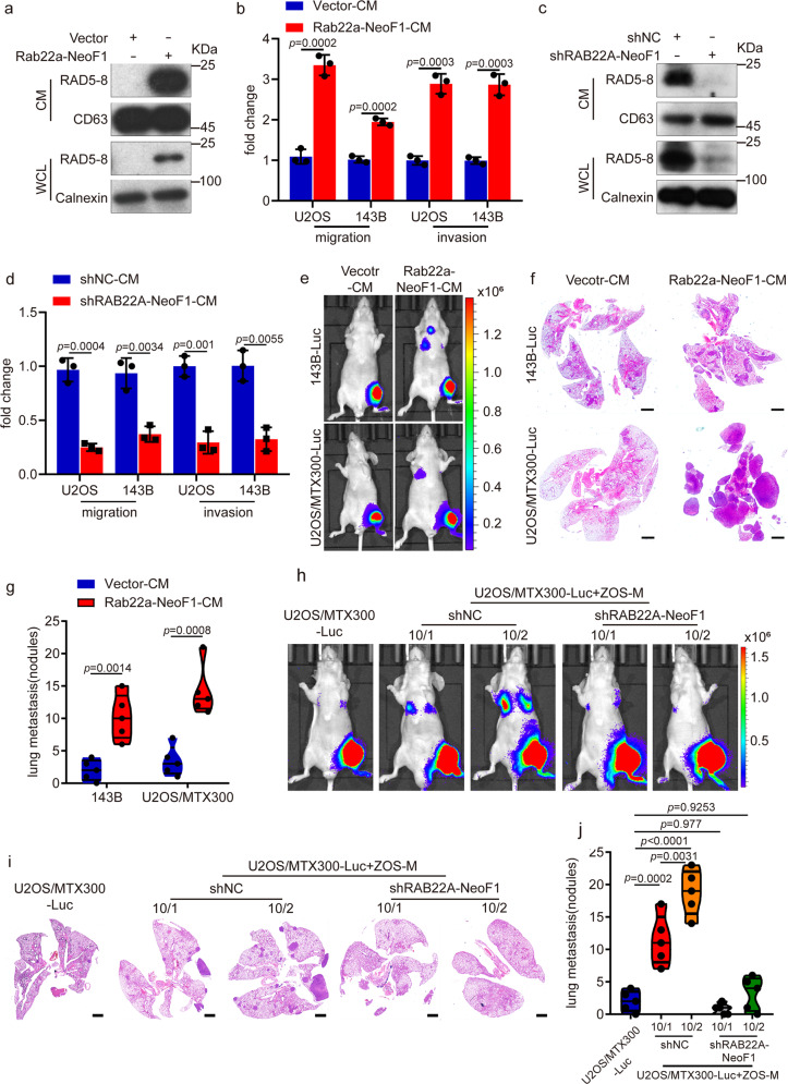
Fig. 1.
The secreted Rab22a-NeoF1 fusion protein promotes metastasis in osteosarcoma. a, c The conditioned media (CM) and cell lysates (WCL) derived from the indicated stable 143B cells (a) and ZOS-M cells (c) were prepared and analyzed by Western blotting. Data in a, c are representative of n = 3 biologically independent experiments. b, d The indicated cells were treated with the conditioned media derived from vector cells (vector-CM), Rab22a-NeoF1 cells (Rab22a-NeoF1-CM), ZOS-M-shNC cells (shNC-CM), or ZOS-M-shRAB22A-NeoF1 cells (shRAB22A-NeoF1-CM) for 24 h and then were subjected to migration and invasion assays. Data are mean ± s.d. of n = 3 biologically independent experiments. P values are shown. e–g Representative IVIS imaging (e), H&E-stained lung sections (f), and quantification of lung metastatic foci (g) from mice orthotopically injected with the indicated cells under treatment of either Vector-CM or Rab22a-NeoF1-CM. n = 5 biologically independent mice. Data are mean ± s.d. P values are shown. Scale bar, 2 mm. h–j Representative IVIS imaging (h), H&E-stained lung sections (i), and quantification of lung metastatic foci (j) from mice orthotopically co-injected U2OS/MTX300-Luc cells with either ZOS-M-shNC cells or ZOS-M-shRAB22A-NeoF1 cells at the indicated ratios. n = 5 biologically independent mice. Data are mean ± s.d. P values are shown. Scale bar, 2 mm

