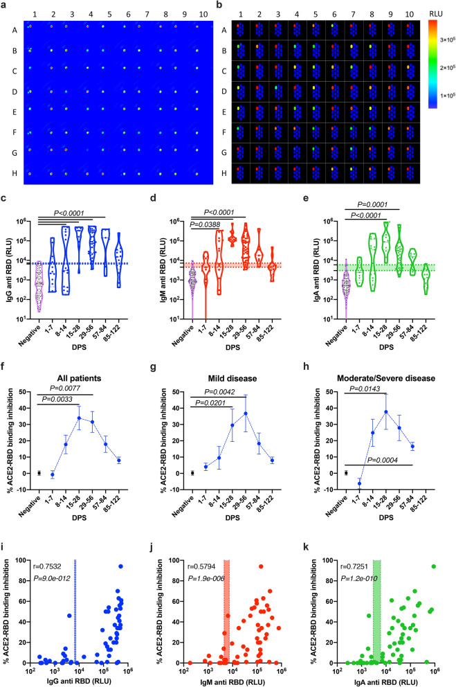
Figure 4.
Anti-SARS-CoV-2-RBD neutralization antibodies using spotted electrochemiluminescence ELISA. Peripheral blood was collected from hospitalized COVID-19 and anonymous recovered patients (n = 75). Negative samples were obtained from true SARS-CoV-2 negative patients (i.e., prior to the SARS-CoV-2 pandemic) (n = 4). Plasma was obtained, diluted 1:50, and added to a 10-spot 96-well plate spotted with SARS-CoV-2 RBD antigen on spot number 1, and BSA on spots number 2–10. ACE2-sulfotag was used instead of a secondary/detection antibody. Representative photomicrographs of real time (a) and schematic (b) images of the spotted 96 well plate are presented. Kinetics of all patients (c), mild (d) and moderate/severe (e) patients is shown; average ± SEM. Inhibition of ACE2-RBD binding was calculated from the average RLU of the 4 negative plasma donors. (f–h) Patients’ antibody results were graphed against the neutralization antibody response. (i–k) Correlation analysis of antibody vs. ACE-RBD binding. Data were calculated using GraphPad Prism 8; the dotted X-line represents the calculated cutoff values (95% and 98% sensitivity) discriminating between positive and negative samples. (c–e) A Nonparametric Kruskal–Wells test for multiple comparisons. (i–k) Correlation analysis was performed using a nonparametric Spearman’s correlation test (two-tailed, 95% confidence). P values are shown.

