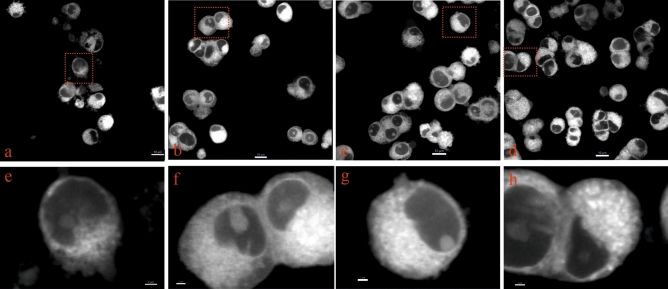
Figure 3.
Changes in mitochondrial morphology in glutamate-exposed PC-12 cells are prevented by ADA-409-052. MitoTracker Red CMXRos was used to stain mitochondria of PC-12 cells after 24 h of exposure to (a,e) 20 mM glutamate, (b,f) glutamate and 10 µM ADA-409-502, or (c,g) ADA-409-052 only, plus control (d,h; media only). (b–d,f–h) Cells, exposed to ADA-409-052 in absence or presence of glutamate show a similar distribution and density of MitoTracker Red CMXRos-positive structures as control cells. (a,e) However, in glutamate-exposed cells appear distribution and density altered, and the overall number of viable cells reduced. Mitochondria were detected by confocal microscopy in live cells and magnified images (e–h) were produced using Imaris; red boxes indicate magnifications. Scale bar: 10 µm; 2 µm for magnifications.

