FIGURE 2.
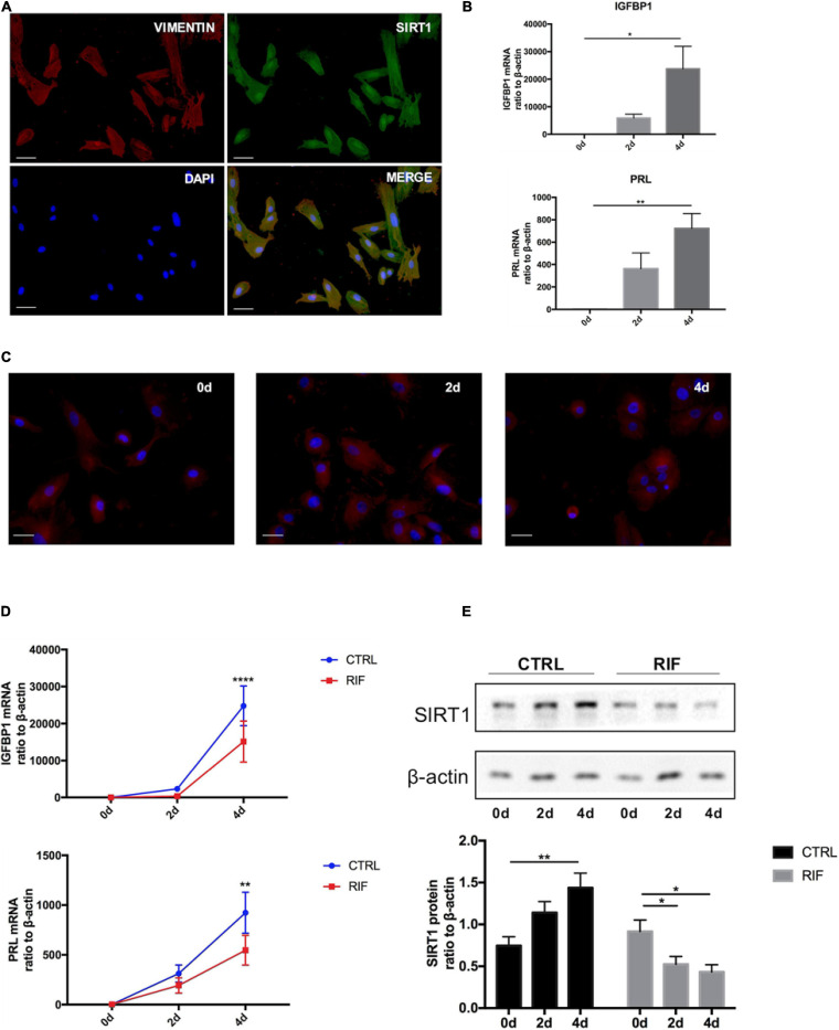
The changes of SIRT1 in decidualization of human ESC. (A) Immunofluorescence staining of SIRT1 (green) and vimentin (red) were performed human ESCs. The nuclear were stained with DAPI (4′,6-diamidino-2-phenylindole) (blue). Scale bar is 50 μm. (B) The mRNA levels of PRL and IGFBP1 during decidualization (0, 2, and 4 days) in vitro. mRNA expression levels were normalized to β-actin. Bar graph was the average data of three independent experiment. (C) A cytoskeletal staining of α-tubulin (red) demonstrated differences in cell shape and cytoskeletal architecture. The nuclear were stained with DAPI (4′,6-diamidino-2-phenylindole) (blue). Scale bar is 50 μm. (D) The mRNA levels of IGFBP1 and PRL in ESC during decidualization in vitro from CTRL (n = 7) and RIF (n = 7). (E) Western blotting analysis of SIRT1 protein abundance in ESC during decidualization in vitro from CTRL (n = 7) and RIF (n = 7), blots graph was representative and bar graph was the average data. *P < 0.05, **P < 0.01, and ****P < 0.0001 vs. 0 days or CTRL (data are means ± SEM).

