FIGURE 3.
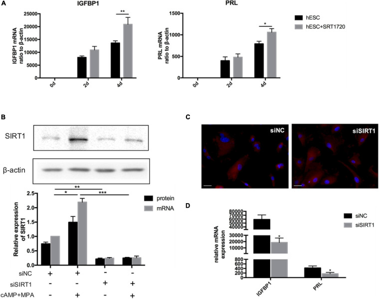
The effect of SIRT1 on ESCs decidualization. (A) The mRNA levels of IGFBP1 and PRL following addition of SRT1720 (10 μM) during decidualization (0, 2, and 4 days) in vitro. (B) Efficiency of siRNA-mediated knockdown in SIRT1 protein abundance and mRNA level. Blots are representative and the bar graphs are the average data of four independent experiment. (C) A cytoskeletal staining of α-tubulin (red) demonstrated cellular morphology after 4-day induced decidualization between siNC and siSIRT1 groups. The nuclear were stained with DAPI (4′,6-diamidino-2-phenylindole) (blue). Scale bar is 50 μm. (D) The mRNA levels of decidual biomarkers after 4-day induced decidualization between siNC and siSIRT1 groups. mRNA expression levels were normalized to β-actin. Bar graph was the average data of four independent experiments. *P < 0.05, **P < 0.01, and ***P < 0.001 (data are means ± SEM).

