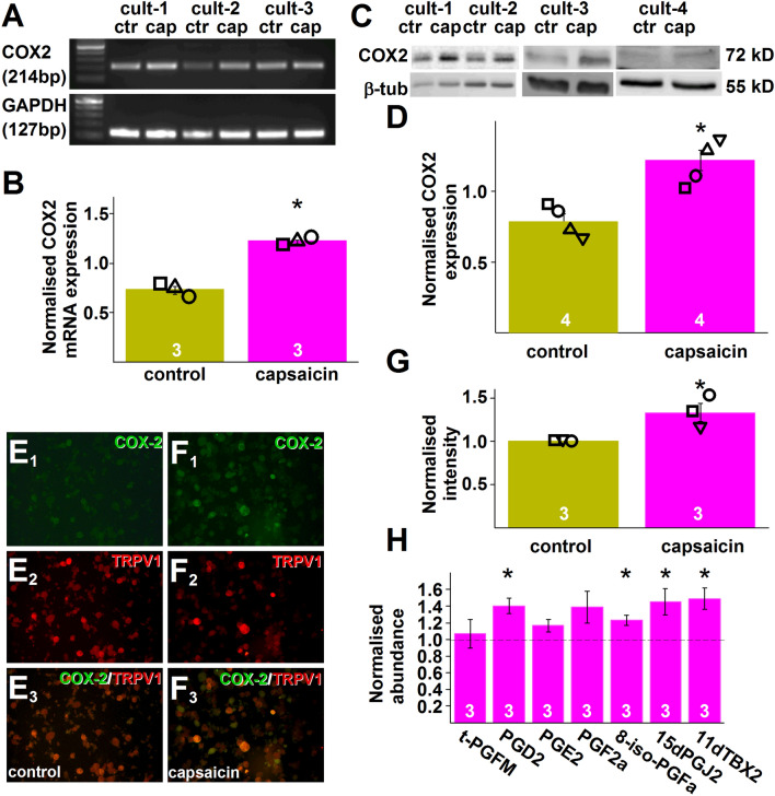
Figure 2.
Capsaicin application to cultured murine primary sensory neurons upregulates COX2 expression and synthesis of a series of COX products. (A) Gel image of RT-PCR products amplified using primer pairs for murine ptgs2 (COX2) and gapdh (GAPDH) and cDNA prepared using RNA isolated from three cultures (cult-1–cult-3) of murine primary sensory neurons 25 min after incubating the cells in control (ctr) buffer or in the presence of 500 nM capsaicin (cap) for 5 min. (B) Averaged normalised ptgs2 expression in cultures shown in (A). Capsaicin (500 nM, capsaicin) application resulted in a significant increase in ptgs2 expression (p = 0.0004; Student’s t-test, n = 3). (C) Gel images of immunoblots prepared using proteins extracted from four cultures (cult-1–cult-4) of murine primary sensory neurons 25 min after incubating the cells in control (ctr) buffer or in the presence of 500 nM capsaicin (cap) for 5 min and antibodies raised against COX2 and GAPDH. (D) Averaged normalised COX2 expression in cultures shown in (C). Capsaicin (500 nM, capsaicin) application resulted in a significant increase in COX2 expression (p = 0.0025; Student’s t-test, n = 4). (E1,E2,E3,F1,F2,F3) Microscopic images of cultured murine primary sensory neurons incubated in control buffer (E1,E2,E3) or in the presence of 500 nM capsaicin (F1,F2,F3) for 5 min. Cultures were fixed 25 min after the incubation and incubated in anti-COX2 (green) and anti TRPV1 antibodies (red). (E1,F1) show COX2 staining, (E2,F2) show TRPV1 staining whereas (E3,F3) show composite image. Note that COX2 staining intensity is increased in the culture incubated in the presence of capsaicin. (G) Normalised averaged intensity values of COX2 immunostaining in three cultures of murine primary sensory neurons. Capsaicin significantly increased the staining intensity (p = 0.0337; Student t-test; n = 3). (H) Targeted UPLC-MS measurements of seven COX products in an eicosanoids panel using samples prepared from cultured murine primary sensory neurons incubated in control buffer or 500 nM capsaicin for 5 min. Samples were prepared 25 min after finishing the 5 min incubation. During the 25 min, cells were kept in the presence of the COX2 substrate arachidonic acid (10 μM). Four of the seven COX products present in the eicosanoid panel exhibited significant increase by incubating the cells in capsaicin (PGD2: q = 0.032; 8-iso-PGF2α: q = 0.028; 15dPGJ2: q = 0.046; 11dTXB2: q = 0.031; Student-t test followed by Benjamini–Hochberg FDR, n = 3).

