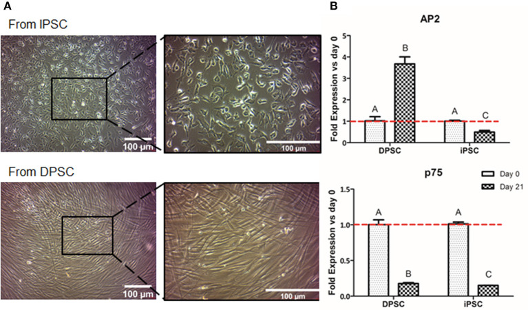
Figure 3.
Formation of NCSC using an adherent method. Microscope images of NCSC-derived from iPSC and from DPSC at day 21 of differentiation (A). Relative mRNA expression in NCSC-derived from DPSC and iPSC, at days 0 and 21, analyzed by quantitative RT-PCR (B). *Scale bar: 100 μm. **Data presented are means ± SD, n = 3 technical replicates per experimental group. Different letter denotes significant differences (p < 0.05). Same letter denotes non-significant differences.

