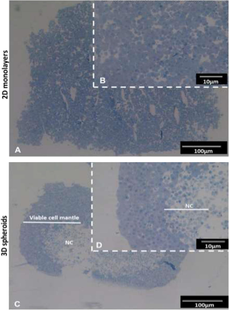
FIGURE 1.
Histological images of INS-1 2D monolayers and 3D spheroid semi-thin sections stained with toluidine blue. Sections of 2D monolayers revealed morphological features such as euchromatic nucleic and prominent nucleoli typical of protein producing cells (A,B). 3D spheroids displayed an outer mantel of viable cells with euchromatic nuclei prominent nucleoli and a central necrotic core (NC) (C,D). Sections captured using 10× and 40× objectives.

