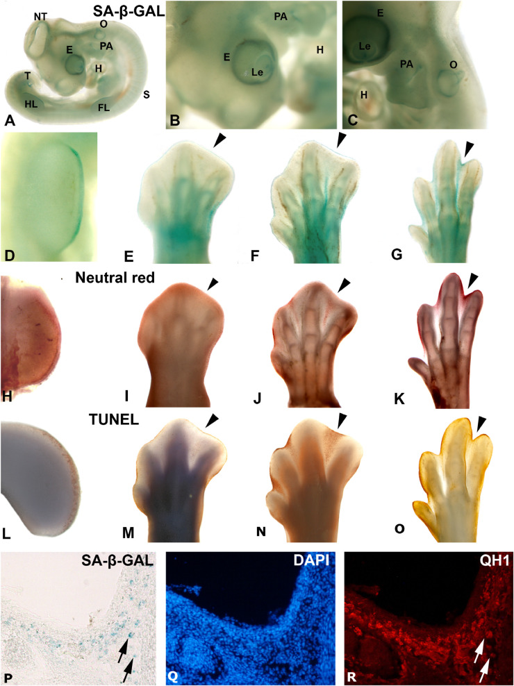
FIGURE 1.
Areas segmenting SA-β-GAL activity and apoptosis during avian embryonic development. Detection of SA-β-GAL activity in embryonic day 3.5 (A,B) and E4 (C) chicken embryos, and E3.5 (D), E6 (E), E7 (F), and E8 (G) hindlimbs. Neutral red staining for cell death detection in E3.5 (H), E6 (I), E7 (J), and E8 (K) hindlimbs. TUNEL assay for apoptosis detection in E4 (L), E6 (M), E7 (N), and E8 (O) hindlimbs. Labeling of AER can be noted in (D,H, and L). Arrowheads in (E–G), (I–K), and (M–O) point to the third interdigital space during the establishment of cell senescence and the progression of interdigital programmed cell death. SA-β-GAL histochemistry (P) and QH1 immunostaining (R) label macrophages in the interdigital mesenchyme of quail at stage 36. DAPI staining (Q) shows the structure of the interdigital space. E, eye; Fl, forelimb; H, heart, HL, hindlimb, Le, lens; NT, neural tube, O, otic vesicle; PA, pharyngeal arches; T, tail bud.

