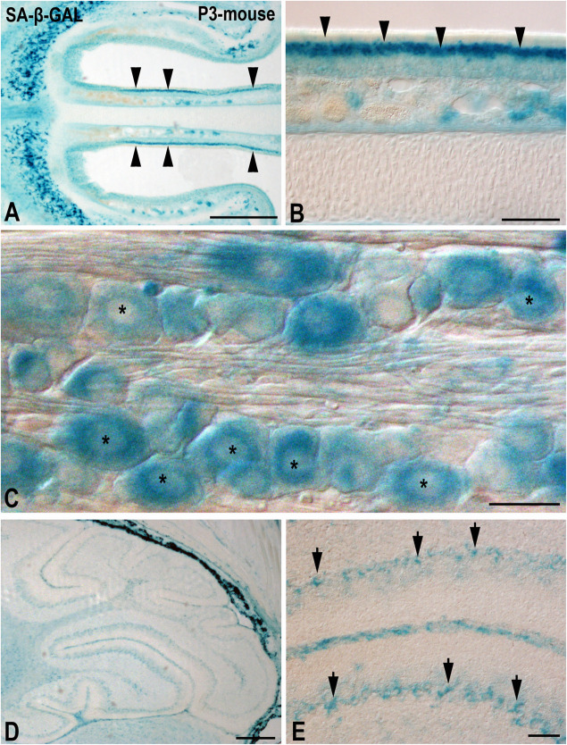
FIGURE 2.
The presence of SA-β-GAL activity in the postnatal day P3 mouse head tissue. Horizontal (A,B) and sagittal (C–E) cryosections were treated with SA-β-GAL histochemistry. (A,B) Intense SA-β-GAL signal is found in the intermediate layer of the olfactory epithelium (arrowheads). (C) Strong SA-β-GAL staining is detected in sensory neurons in the trigeminal ganglion (asterisks). (D,E) SA-β-GAL activity is detected in the cerebellum, mainly in the Purkinje cell layer (arrows). Scale bars: 200 μm (A,D), 50 μm (B,E), and 20 μm (C).

