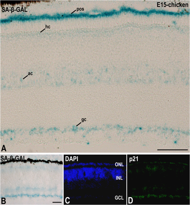
FIGURE 3.
The presence of SA-β-GAL activity in the embryonic day E15 chicken retina. Cryosections of retinas were treated with SA-β-GAL histochemistry (A,B) and antibodies against p21 (B–D). DAPI staining shows the laminated structure of the retina (C). SA-β-GAL staining is found in the photoreceptor outer segments and in subpopulations of amacrine and ganglion cells (A,B). The horizontal cell layer appears faintly labeled (A,B). p21 immunostaining strongly correlates with the SA-β-GAL labeling pattern. ac, amacrine cells; gc, ganglion cells; GCL, ganglion cell layer; hc, horizontal cells; INL, inner nuclear layer; ONL, outer nuclear layer; pos, photoreceptor outer segments. Scale bars: 50 μm (A and B–D).

