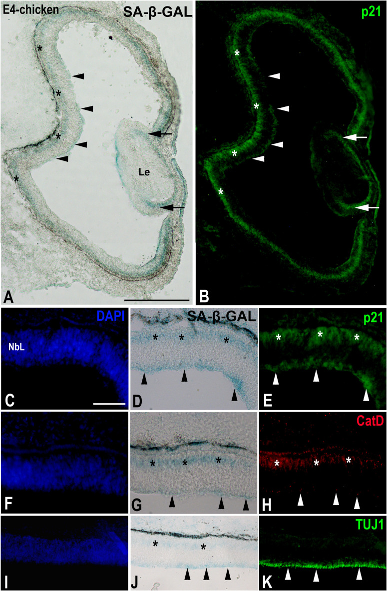
FIGURE 4.
The presence of SA-β-GAL activity in the embryonic day E4 chicken retina. Cryosections of retinas were treated with SA-β-GAL histochemistry and antibodies against p21 (A–E), CatD (F–H), and TUJ1 (I–K). DAPI staining shows that the neural retina consists of a NbL (C,F,I). SA-β-GAL staining is detected in the scleral (asterisks in A,D,G,J) and vitreal (arrowheads in A,D,G,J) regions of the retina. p21 immunostaining correlates with the SA-β-GAL staining pattern in the undifferentiated retina (arrowheads and asterisks in B,E) and lens (arrows in B). CatD immunoreactivity (arrowheads and asterisks in H) is strongly coincident with the SA-β-GAL histochemistry signal (arrowheads and asterisks in G). TUJ1 immunoreactivity is intense in the vitreal surface of the NbL (arrowheads in K), coinciding with the vitreal SA-β-GAL histochemistry signal detected in the same region (arrowheads in J). Le, lens; NbL, neuroblastic layer. Scale bars: 150 μm (A,B), 50 μm (C–K).

