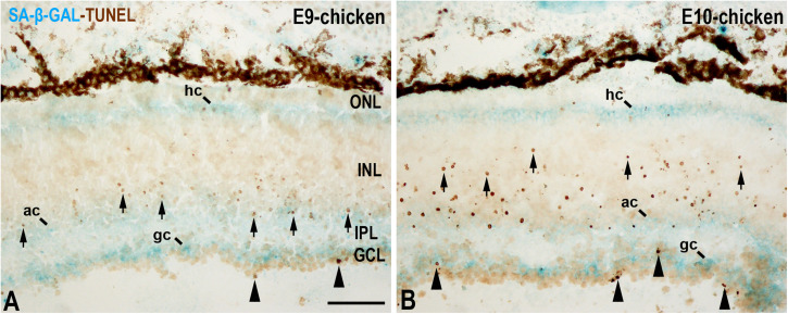
FIGURE 5.
SA-β-GAL activity and cell death in the embryonic day E9 (A) and E10 (B) chicken retina. Cryosections were doubly stained with SA-β-GAL histochemistry and TUNEL technique. TUNEL-positive nuclei are mainly detected in the GCL (arrowheads) and in the middle region of the INL (arrows). SA-β-GAL activity is observed in the GCL, amacrine cell layer, and horizontal cell layer. ac, amacrine cells; gc, ganglion cells; GCL, ganglion cell layer; hc, horizontal cells; INL, inner nuclear layer; IPL, inner plexiform layer; ONL, outer nuclear layer; pos, photoreceptor outer segments. Scale bar: 50 μm.

