Figure 3.
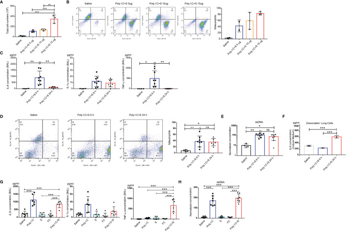
Polyinosinic-polycytidylic acid (poly[I:C]) + recombinant SARS-CoV-2 spike-extracellular domain protein (SP)-induced cytokine-release storm in the mouse lung. (A) Total cell count in the BAL from poly(I:C) + 5-, 10-, or 15-µg SARS-CoV-2 spike protein-challenged mice; control group, Saline (each n ≥ 3); (B) Cellular composition in BAL. (C) Production of IL-6, IL-1α, and tumor necrosis factor (TNF)α in the bronchoalveolar lavage fluid (BAL) from poly(I:C) + SP-challenged mice. 6 or 24 h after SARS-CoV-2 mimic challenged mice. Control group; Saline (each n ≥ 4); (D) percentage of neutrophils in BAL; (E) Levels of dsDNA in the BAL. Single cells dissociated from the lung tissue of poly(I:C) + SP-challenged mice. (F) IL-6 concentration from cell culture medium. (G) Concentrations of IL-6, IL-1α, and tumor necrosis factor (TNF)α in bronchoalveolar lavage fluid (BALF) after challenge with SARS-CoV-2 mimic (poly[I:C] + SP), poly(I:C) alone, SP alone, FC alone, saline control (each n ≥ 5), (H) Levels of dsDNA in the BALF. *P < 0.05; **P < 0.01; ***P < 0.001, ns (non-significant), p > 0.05, one-way ANOVA with a post hoc Bonferroni test.

