
Fig. 1.
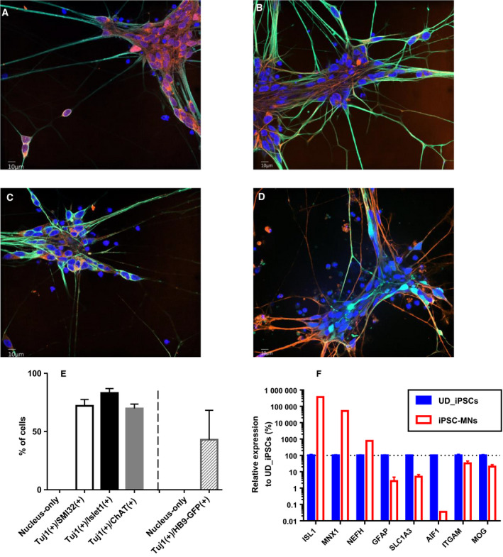
Preparation of human iPSC‐derived motor neurons. Human iPSCs were differentiated to motor neurons. (A–C) Green and blue colors represent the immunostaining of Tuj1 and DRAQ7‐stained cellular nuclei, respectively. Orange color represents Islet1 (A), SMI‐32 (B) and ChAT (C), respectively. The image was obtained at day 7 after seeding of MNPs at original magnification of 60×. Scale bars: 10 μm. (D) Green, orange and blue colors represent the fluorescence of eGFP, immunostaining of Tuj1 and NucSpot Live 650‐stained cellular nuclei, respectively. The image was obtained at day 7 after seeding of MNPs at original magnification of 60×. Scale bar: 10 μm. (E) Purity of each population was analyzed according to the method described in the Materials and Methods. The average of six replicated wells is represented. The error bar indicates SD of n = 6. (F) The expression levels of cell‐type‐specific genes were investigated by RT‐qPCR and normalized by ACTB. Those in 201B7 iPSCs‐derived motor neurons (MNs) were compared with those in undifferentiated (UD) iPSCs. The average of three replicates is represented, and the bar indicates SD of n = 3.