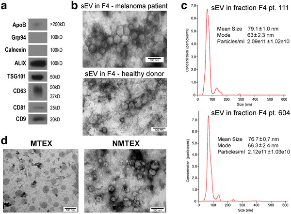
FIGURE 1.

Characterization of sEV isolated from plasma. Panel a – western blot characterization of exosome markers in sEV collected in fraction #4 as described in Materials and Methods. Panel b – a TEM image of sEV in fraction #4 obtained from plasma of a melanoma patient or healthy donor. Panel c – NanoSight profiles of EVs in fraction #4 of two patients with melanoma. Panel d – TEM of MTEX detached from anti‐CSPG4 mAbs on beads and NMTEX that remain in suspension following immune capture
