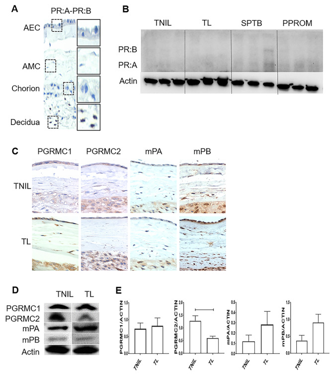
Figure 1.

Differential expression of P4 membrane receptors in human fetal membranes at term. (A) Fetal membrane cells, including amnion epithelial cells (AECs), amnion mesenchymal cells (AMCs), and chorion cells, were negative for nuclear progesterone receptors A and B (PR:A and PR:B) (×40). Immunohistochemistry localized PRA and PRB in the maternal decidua, confirming the specificity of staining. (B) Western blot analysis of nuclear P4 receptors (PR:A–PR:B) in term labor (TL), term not in labor (TNIL), and preterm birth fetal membranes (amnion–chorion). (C) All four P4 membrane receptors, progesterone receptor membrane component (PGRMC) 1, PGRMC2, mPα, and mPβ, in fetal membrane cells were localized by immunohistochemistry at term, regardless of the labor status (×40) (N = 3); Scale bar = 50 μm. (D) Western blot analysis of membrane P4 receptors at TL and TNIL in fetal membranes (amnion–chorion). (E) Densitometry analysis of TL showed significantly lower expression of PGRMC2 compared with TNIL sample (P = 0.01), whereas PGRMC1, mPα, and mPβ expression did not change in TL compared with TNIL fetal membranes (N = 5; mean ± SEM). Full gels for (C) can be found in Supplementary Figure 2. PGRMC1 and PGRMC2 data also seen in Richardson et al. [12].
